
No single test can accurately diagnose cancer. Many diagnostic tests are required to determine whether the patient has cancer. If you show any symptoms, the doctor will conduct a series of screening tests to decide whether or not it is due to cancer or some other cause.
Effective diagnostic tests confirm or eliminate the presence of diseases. They also monitor the disease process and plan for and evaluate the effectiveness of treatment.
Sometimes, it is necessary to repeat some tests when a patient's condition has changed. This may happen if a sample collected needs to be of better quality or in cases where an abnormal test result has to be confirmed.
Get in Touch with Medical Experts
The diagnostic procedures for cancer may include the following:
- Imaging [mammograms, ultrasounds, and magnetic resonance images (MRI), X-rays, computed tomography (CT) scans, and fluoroscopy, PET CT scan]
- Endoscopic examination
- Laboratory tests (including blood tests and tests for tumor markers)
- Biopsy (Endoscopic biopsy, bone marrow biopsy, excisional or incisional biopsy, fine needle aspiration biopsy, punch biopsy. shave biopsy, skin biopsy, etc.)
- Genetic testing
Here we will discuss some basic tests that are used to help diagnose cancer. Depending on the symptoms, some patients may have other tests too.
Which Tests are Performed for Cancer Diagnosis?
The specialist may start by getting details about your family and medical history. To diagnose cancer, the doctor may conduct a physical exam, order lab tests, imaging tests (scans), biopsy, or other tests or procedures.
1. Colonoscopy
A colonoscopy exam is performed for changes such as swollen, irritated tissues, polyps, or cancer in the large intestine (colon) and rectum.
How is it performed?
- A flexible long tube known as a colonoscope is inserted into the rectum.
- The doctor views the inside of the colon with the help of a tiny video camera at the tip of the tube.
- If necessary, polyps or other abnormal tissues can be removed during a colonoscopy.
2. Imaging Tests
a. Mammogram - This is an X-ray image of the breast used to detect early indications of breast cancer.
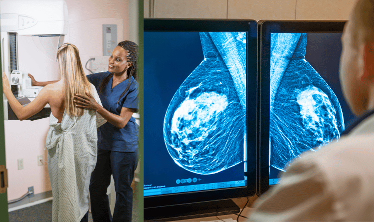
How is it performed?
- During a mammogram, an X-ray captures black-and-white images.
- These images, displayed on a computer screen
b. Breast MRI - A breast MRI is performed after you have a positive biopsy for cancer.
How is it performed?
- The breast MRI captures multiple images of your breasts to create a detailed picture.
- This picture shows where tumors may exist.
3. Tumor Marker Blood Tests
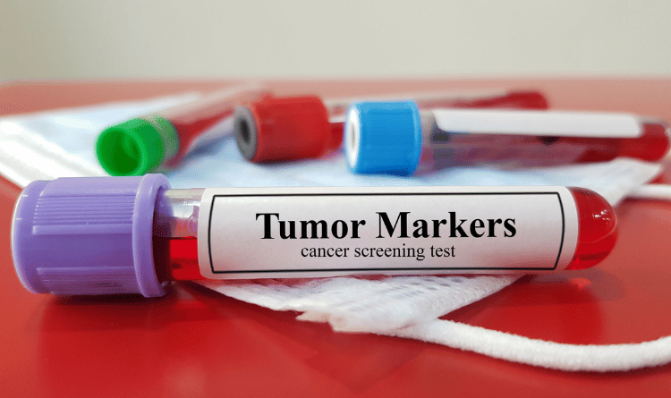
a. Cancer Antigen (CA)-125 - This test measures the amount of the protein CA-125 in the blood. The test can be utilized for monitoring certain cancers before and after treatment. Additionally, it may be employed to detect early signs of ovarian cancer in individuals at a very high risk of developing the disease.
How is it performed?
- A blood sample, collected by inserting a needle into a vein, usually in the hand or arm, is sent to a lab for analysis.
b. CA 15-3 - CA 15-3 is a substance that stimulates your body's defense system. Some cancer cells release the CA 15-3 antigen into the blood. This test monitors certain types of cancer, such as breast cancer.
How is it performed?
- A blood sample, collected by inserting a needle into a vein, usually in the hand or arm, is sent to a lab for analysis.
c. CA 27-29 - This test measures how much CA 27-29 is in your blood. Antigens like CA 27-29 give information about tumor markers.
How is it performed?
- A blood sample, collected by inserting a needle into a vein, usually in the hand or arm, is sent to a lab for analysis.
d. Carcinoembryonic antigen (CEA) - CEA protein is typically produced by cancerous cells and may also be produced with conditions such as liver disease and inflammatory bowel disease. Since CEA may be detected in the blood or bodily fluids when cancer is present, the test may help determine whether the patient has this disease. Typically, this test is used to monitor colon, rectum, liver, breast, pancreas, and ovarian cancers.
How is it performed?
- A blood sample, collected by inserting a needle into a vein, usually in the hand or arm, is sent to a lab for analysis.
e. Prostate-Specific Antigen (PSA) Test - Both normal and cancerous cells in the prostate gland produce PSA protein. A blood test is used to measure the amount of PSA present in the bloodstream.
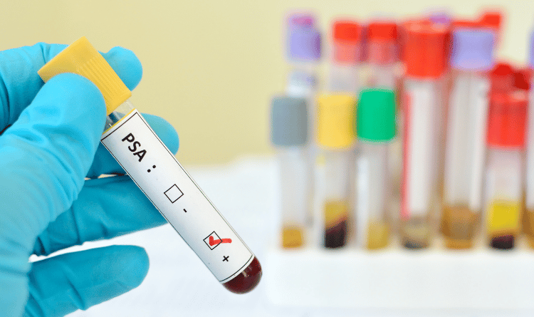
How is it performed?
- A blood sample, collected by inserting a needle into a vein, usually in the hand or arm, is sent to a lab for analysis.
f. Human epidermal growth factor receptor PN2 (HER2) protein - HER2-positive test determines protein HER2, which promotes the growth of cancer cells.
How is it performed?
- HER2 status testing is performed on a biopsy sample taken from the tumor.
- The two ways to test HER2 status include Immunohistochemistry (IHC) and Fluorescence in situ hybridization (FISH).
g. Neurotrophic tyrosine receptor kinase (NTRK) fusion genes - NTRK gene fusions are biomarkers for cancer therapy. These can be found in over 25 types of cancer, regardless of their location.
How is it performed?
NTRK gene fusions biomarker tests may include:
- Next-generation sequencing (NGS)
- Immunohistochemistry (IHC)
- DNA fluorescence in situ hybridization (FISH)
- Polymerase chain reaction (PCR)
h. Programmed Death Ligand (PD-L1) Protein - A test for PD-L1 involves analyzing a tissue sample from a cancerous tumor to determine the amount of PD-L1 protein in the cancer cells. If you have certain types of cancer, PD-L1 testing can check whether you may benefit from a cancer treatment called immunotherapy.
How is it performed?
- The test is done on a tissue sample from a tumor.
- If you're having surgery to remove a tumor, a sample will be taken for testing.
- If you aren't having the tumor removed, you may have a biopsy.
i. Estrogen Receptor (ER) and Progesterone Receptor (PR) - ER and PR test helps determine the patient's risk of recurrence. It also determines the type of treatment most likely to lower the risk of recurrence.
How is it performed?
- ER/PR testing is done using a sample of tissue taken during a biopsy. This same tissue sample is also used to diagnose cancer.
- The ER/PR test results help doctors plan treatment and understand how likely the cancer is to grow or spread.
j. MSI and MMR testing - MicroSatellite Instability (MSI) and MisMatch Repair (MMR) are both biomarkers commonly tested to identify whether immunotherapy is likely to work in cancer when cells in the body divide and any errors in the genetic makeup of the cells are repaired.
How is it performed?
- MSI is a Polymerase Chain Reaction (PCR) test that compares tumor tissue to normal tissue to measure microsatellite instability.
- MMR is an Immunohistochemistry (IHC) test tested on the tumor tissues of cancer or when the cancer tissue is surgically removed for mismatch-related proteins such as MLH1, MSH2, MSH6, and PMS2.
- Usually, these proteins are present, and if they are not expressed in the IHC test, then it is deficient MMR.
4. Genetic and genomic testing

a. Breast Cancer 1 (BRCA1) and Breast Cancer 2 (BRCA2) mutations test - This is a blood test that uses DNA analysis to identify mutations in either of these two breast cancer susceptibility genes.
How is it performed?
- A BRCA gene test uses a blood sample, saliva (spit), or cells inside your cheek to look for changes in your BRCA 1 and BRCA 2 genes that may increase your risk of cancer.
b. Estrogen Receptor 1 (ESR1) gene mutations - These tests assist in the clinical management of metastatic breast cancer patients. It identifies tumors with evolving resistance to endocrine therapy.
How is it performed?
- This evaluation utilizes specialized next-generation sequencing to detect somatic mutations in the ESR1 gene. Its purpose is to identify somatic mutations in solid tumor samples, not to examine any germline alterations in the ESR1 gene.
- The tests detect the somatic mutations in the ESR1 gene using targeted next-generation sequencing. It is specifically designed to assess solid tumor samples and does not test for germline alterations in the ESR1 gene.
c. Phosphatidylinositol-4,5-bisphosphate 3-kinase catalytic subunit alpha (PIK3CA) gene mutation - The PIK3CA family of enzymes plays a vital role in functions such as cell growth, proliferation, motility, differentiation, survival, and intracellular trafficking. These functions are closely associated with cancer.
How is it performed?
- Plasma or tissue specimens can be used to detect PIK3CA mutations.
- Your doctor will send a tissue or blood sample (to get a plasma specimen).
- If you've had a biopsy done before, ask your doctor if your tissue is still viable for PIK3CA gene mutation testing.
- If you do not, your doctor may start with a blood test. If no mutation is found in the plasma specimen, ask your doctor to test your tissue to confirm.
How does the Cost of Cancer Diagnostic Tests Vary in Different Countries?
Cancer diagnostic tests are crucial in early detection and treatment planning, significantly impacting patient outcomes. The cost of these tests may vary widely across different countries, particularly in regions like India, Thailand, and Turkey.
In India, where healthcare accessibility is a concern, the cost of cancer diagnostic tests can be comparatively lower than in many Western countries. This is due to affordable medical facilities and a highly competitive diagnostic market.
Thailand offers a blend of affordability and quality. The cost may be more reasonable than equivalent services in countries like the United States. Similarly, Turkey provides cancer diagnostic tests at a relatively lower cost than developed nations. Patients can access various tests, from basic screenings to advanced molecular diagnostics, at a fraction of the cost in some Western countries.
It is essential to consider the entire spectrum of cancer care and consult with medical professionals before making decisions about diagnostics and treatment.
To Conclude
If the biopsy and other tests indicate the presence of cancer, additional examinations may be necessary to aid your doctor in planning your treatment. Your doctor may conduct further tests to determine if the cancer has spread and to what extent, as this data is crucial in determining the stage of your cancer.
In some instances, additional pathology studies may be conducted to determine the grade of the tumor or tumor markers may be examined to ascertain your risk group. This information is crucial in determining the most effective treatment plan. Furthermore, your tumor may undergo additional testing to identify other tumor markers or biomarkers.


 in Germany: Understanding the Procedure")


